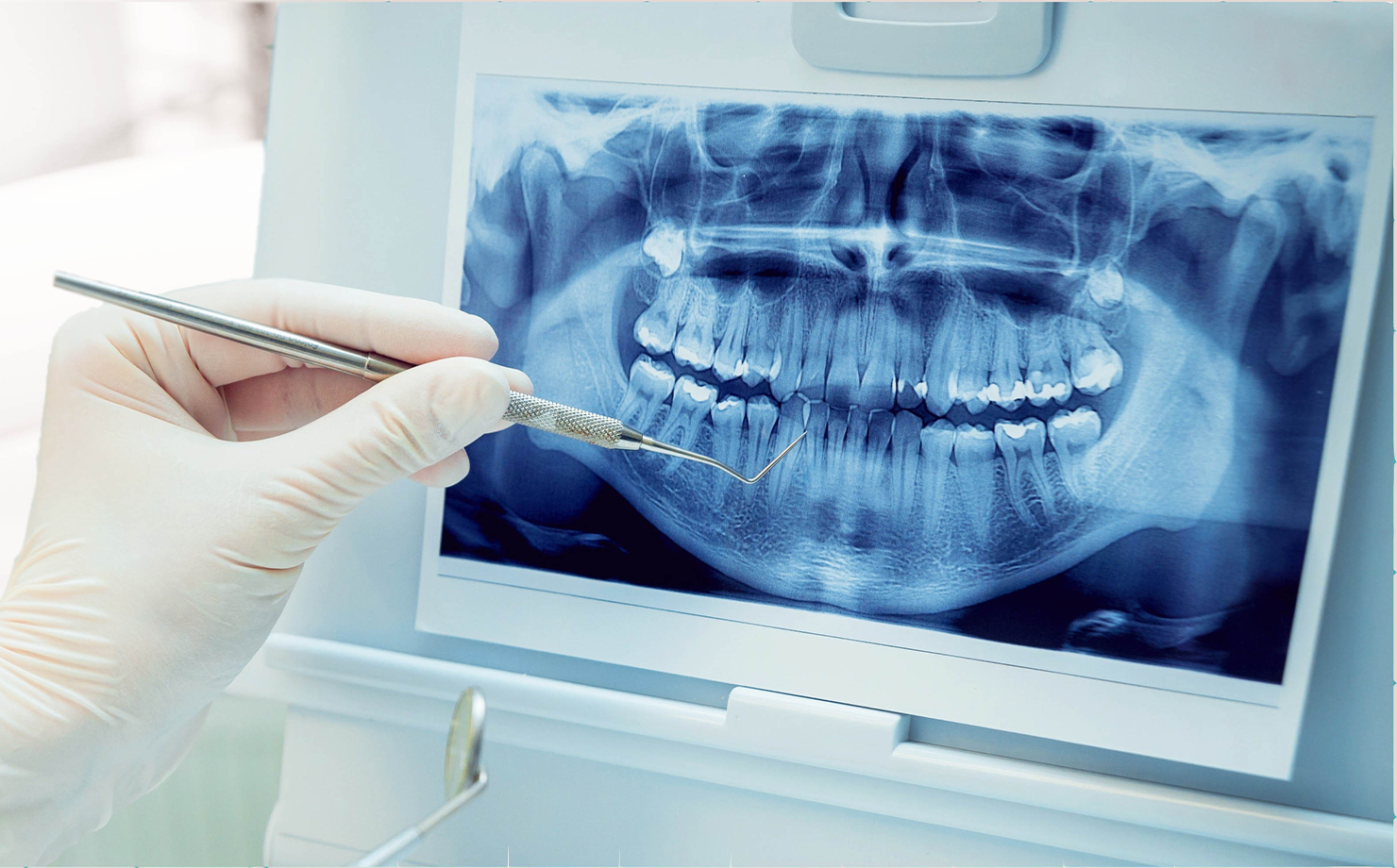
Proper treatment begins with an accurate diagnosis, and the eye of diagnosis is radiology. Unseen parts of the teeth and jawbone can only be clearly evaluated through imaging techniques. How deep a cavity has extended, whether the bone is suitable for an implant, the position of an impacted tooth, or the boundaries of an infection—all of these are revealed through radiological images. Modern digital systems deliver this information with low radiation exposure and high resolution.
What is Oral and Maxillofacial Radiology?
Oral and maxillofacial radiology encompasses the evaluation of teeth, jaws, and surrounding tissues using imaging modalities. This field ranges from periapical films showing a single tooth to panoramic X-rays offering a complete view of both jaws in a single frame, as well as 3D dental tomography. Compared to traditional film, digital imaging provides significantly lower radiation doses and immediate results. These images serve as the foundation of your treatment plan.
Who is it Suitable For?
Almost every patient visiting for a comprehensive examination benefits from a radiological evaluation based on their needs. Three-dimensional imaging is particularly valuable for patients undergoing implant planning, those with impacted teeth, or individuals requiring oral surgery. In cases involving hidden decay, tooth root and bone issues, or suspected cysts and infections, imaging clarifies the diagnosis. It is also used to monitor dental development in children.
How is it Applied?
The appropriate imaging technique is determined by your dentist according to your specific needs. Periapical X-rays focus on a single area, while panoramic X-rays capture the entire jaw and all teeth in a single pass; these procedures take only seconds. Three-dimensional tomography is performed as the device rotates around the head to capture cross-sectional images of the bone and tissues. All processes are fast, painless, and fully digital. Images are immediately rendered on-screen and evaluated by your physician.
Advantages
Radiological imaging ensures accurate diagnosis by uncovering hidden problems early. Digital systems operate with low radiation doses and provide instant results. Three-dimensional imaging enhances treatment safety by delivering millimeter-precision accuracy, particularly in implant placement and surgical planning. Storing images digitally facilitates comparative tracking throughout the treatment journey. Proper planning prevents unnecessary procedures and unexpected surprises.
Treatment Process
Radiological imaging is usually conducted during the same appointment as part of the initial examination. Depending on the type of image required, the process takes from a few seconds to a couple of minutes. Because results are obtained instantly in a digital environment, there is no waiting time; your physician evaluates the images alongside you. In light of the findings, a personalized treatment plan is constructed or existing plans are refined.
Recovery Period
Since radiological imaging is a non-invasive procedure, it requires no recovery period. The process is completely painless, allowing you to return to your daily activities immediately without any restrictions. The minimal radiation dose of digital systems makes the procedure safe. When necessary, images taken at different times are compared to monitor the progression of the treatment.
Frequently Asked Questions
- Are dental X-rays harmful? Radiation doses in digital dental X-rays are extremely low. Your dentist orders imaging only when necessary and to the extent required.
- What is the difference between a panoramic X-ray and 3D tomography? A panoramic X-ray displays the entire jaw in a single two-dimensional frame, whereas tomography provides cross-sectional 3D images for a far more detailed evaluation.
- Is tomography strictly required for dental implants? In most implant planning cases, 3D imaging is of immense value for viewing jawbone volume and structure, ensuring safe planning.
- Can dental X-rays be taken during pregnancy? Unless absolutely necessary, X-rays are postponed during pregnancy. In mandatory cases, they are evaluated with protective measures under doctor supervision; always inform your dentist of your pregnancy.
- When will the imaging results be ready? With digital systems, results appear on the screen instantly; there is no need to wait.
Accurate Diagnosis is the First Step to Effective Treatment
Catching hidden issues early directly impacts treatment success. Modern radiological imaging delivers high-accuracy diagnosis with minimal radiation. Schedule your examination appointment today for a comprehensive evaluation of your oral and dental health.